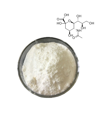
 The Sialic Acid market research report presents valuable information about the industry, including its Compound Annual Growth Rate (CAGR) expressed in USD million, projected up to the year 2030. Furthermore, this report delves into market segmentation driven by key factors and outlines associated business strategies. The Global Sialic Acid Market Report provides Insightful information to the clients enhancing their basic leadership capacity identified with the global Sialic Acid Market business, including market dynamics, segmentation, competition, and regional growth. Report likewise directed a PESTEL analysis in the business to concentrate on key influencers and boundaries to entry. The data from these assays were complemented by a glycan array analysis which determined that AAV1 efficiently binds to NeuAcα2-3GalNAcβ1-4GlcNAc as well as glycoproteins containing NeuAcα2-3/α2-6Galβ1-4GlcNAc N-linked glycans. Taken together, competition, genetic, inhibitor, enzymatic reconstitution, and glycan array experiments support α2,3 and α2,6 sialic acids that are present on N-linked glycoproteins as primary receptors for efficient AAV1 and AAV6 viral infection.
Sialic acids are key structural determinants and contribute to the functionality of a number of immune cell receptors. However, because AAV6 is more efficient than AAV1 for liver transduction (16), this raises the question of how this difference may exist if these serotypes share identical receptors. Finally, we suggest that the immunogenicity of antigen-loaded DCs used in cell vaccines may depend on its sialylation content. To correlate a specific sialylation deficiency with the triggering of DC maturation, we also analysed DCs from ST3Gal.I−/− and ST6Gal.I−/− mice. Evidence from different groups suggests that the state of sialylation of DCs may influence their response.17,18 We have previously studied the surface sialylation of human moDCs and we observed a significantly increased expression of sialylated structures during the differentiation of monocytes into moDCs, most probably as the result of the activity of ST3Gal.I and ST6Gal.I sialyltransferases.19 In addition, we have also observed that the removal of the sialylated structures by neuraminidase treatment diminished the moDC capacity for endocytosis,19 suggesting a triggering of DC maturation. NeuB and NeuC are isolated from bacterial strains that contain sialylated structure in their cells envelope, such as C. jejuni strain ATCC Accession No. 43438. It is also within the scope of the invention to use substantially identical sequences, and/or conservatively modified variations of said sequences as defined hereafter.

 Overall, these data suggest a novel role for the sialylated glycans; in particular, those generated by ST3Gal.I and ST6Gal.I sialyltransferases, in the modulation of the DC maturation state. Human moDCs were generated from monocytes isolated, as described previosuly19, by positive selection using CD14 antibody-coated magnetic beads (Miltenyi Biotech, Bergisch Gladbach, Germany), from peripheral blood mononuclear cells (PBMCs) of healthy volunteers provided and ethically approved by the Portuguese Blood Institute. Real-time PCR was performed in a 7500 Fast Real-Time PCR System (Applied Biosystems) using TaqMan® Fast Universal PCR Master Mix, TaqMan® probes and primers provided by Applied Biosystems. If you liked this write-up and you would certainly such as to get more information concerning manufacturer of sialic acid powder for cosmetic Ingredients kindly browse through the web site. Half of the cells were reserved for RNA extraction and real-time polymerase chain reaction (PCR). The labelled cells were then incubated with the autologous, TT-loaded moDCs (treated or mock-treated with neuraminidase), in the proportion of 8 : 1, in a 96-well round-bottom plate, for 7 days. Particles (2 × 10⁸) of AAV1-luc, AAV4-luc, or AAV6-luc were incubated with the indicated concentrations of mucin for 30 min at 20°C. Virus alone or virus plus mucin were added to Cos-7 cells growing in 24-well plates (2 × 10⁵ cells/well) in equal volumes of DMEM, and cells were incubated for 1 h at 37°C. Cells were rinsed twice with DMEM and incubated at 37°C. Luciferase expression was tested 24 h later.
Cultures were then incubated at 4°C for 20 min, rinsed three times with medium, and then fixed with 4% paraformaldehyde in phosphate-buffered saline (PBS). In these cases, the negative fractions (CD14− PBMCs), obtained after monocyte isolation, were maintained in culture until mixed lymphocyte cultures with autologous moDCs. As a negative control, transduction by AAV2 did not obviously change after resialylation with each sialyltransferase (Fig. (Fig.8C).8C). DC subset. The DCs differentiated in vitro from bone marrow progenitors (BMDCs) of both deficient mouse strains presented increased expression of MHC II molecules and a reduced capacity for endocytosis, when compared with wild-type (WT) mice with normal sialyltransferase expression profiles. The expression of specific maturation markers were examined in ex vivo DCs obtained from blood, lymph nodes and spleen. Cells from spleens and the axillar lymph nodes were obtained by flushing with culture medium and mechanical disruption. 05 mg/ml mitomycin C for 30 min and washed three times with 10 ml RPMI-1640 medium. Tetanus toxoid (TT) and mitomycin C were purchased from Sigma. RPMI-1640 supplemented with 10% fetal calf serum from Sigma (St Louis, MO, USA), 2 mm l-glutamine, 1% non-essential amino acids, 1% pyruvate, 100 μg/ml penicillin/streptomycin and 50 μm 2-mercaptoetanol, all from Gibco-Invitrogen (Paisley, UK) was used throughout this study for cell culture.
